
The relationship between the lymphoid tissues of the pharynx—specifically the tonsils and adenoids—and the mechanics of breathing is far more intricate than a simple physical blockage. While often relegated to the realm of common childhood ailments, the sustained impact of these structures when enlarged, a condition known as hypertrophy, extends into complex physiological and developmental territories. They are, in essence, strategically positioned sentinels in the throat and nasopharynx, guarding the entrance to the respiratory and digestive tracts. Their primary function is immunological, filtering pathogens that are inhaled or ingested, but their location means that any significant increase in volume directly impinges upon the vital passage of air.
The Anatomical Sentry: Tonsils and Adenoids in the Airway Architecture
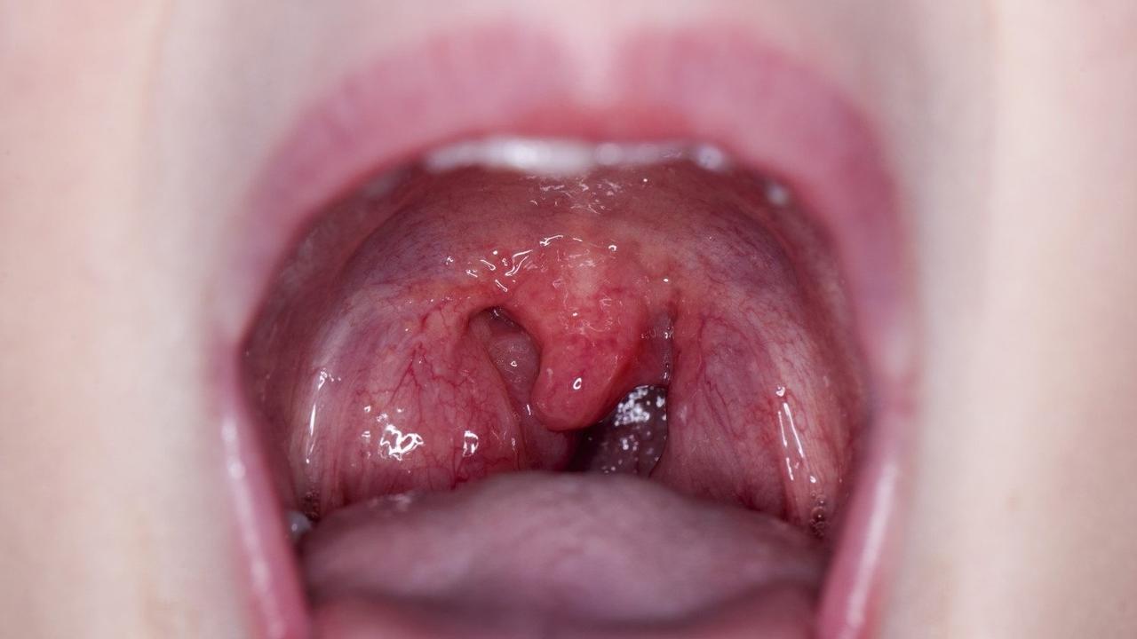
The tonsils, or palatine tonsils, are the two visible masses on either side of the back of the throat, residing in the oropharynx. The adenoids, conversely, are situated higher up, hidden from direct view behind the nose in the nasopharynx. Together, they form part of a defensive ring of lymphoid tissue. In children, particularly between the ages of two and six, these tissues are at their peak of immunological activity and size, making this the critical period for hypertrophy-related breathing issues. The sheer mass of swollen tissue, when disproportionate to the size of the child’s upper airway, becomes a formidable physical obstruction. This is not merely an inconvenience; it represents a significant narrowing of the air corridor that must remain open and compliant during all phases of respiration, especially during sleep.
“The tonsils, or palatine tonsils, are the two visible masses on either side of the back of the throat, residing in the oropharynx.”
The consequences of this obstruction manifest differently depending on which structure is the main culprit. Enlarged adenoids are particularly adept at blocking the nasal passage high up, forcing obligate mouth breathing—a pattern that has its own cascade of physiological and even developmental effects. Conversely, the palatine tonsils, by narrowing the oropharyngeal space, are more frequently implicated in the collapses that lead to frank pauses in breathing during the night. The distinction is crucial for diagnosis and understanding the precise nature of the respiratory disturbance. The chronic inflammatory state, whether driven by frequent infection, environmental allergies, or even a naturally larger size, dictates the degree of compromise to the airway’s patency.
Snoring and Silent Pauses: The Continuum of Sleep-Disordered Breathing
The most noticeable symptom associated with tonsillar and adenoid enlargement is often loud, persistent snoring. This auditory signature is produced by air attempting to rush through a severely constricted passage, causing the surrounding soft tissues to vibrate. Snoring, however, exists on a spectrum of severity that culminates in Obstructive Sleep Apnea (OSA). In OSA, the airway does not just narrow—it completely collapses for brief periods, causing a total cessation of airflow. The body, deprived of oxygen, registers a state of alarm, forcing the individual to briefly wake up, gasp, and restart the breathing cycle.
“The most noticeable symptom associated with tonsillar and adenoid enlargement is often loud, persistent snoring.”
These episodes, known as apneas or hypopneas, may happen scores of times each hour, preventing the brain from achieving the deep, restorative phases of sleep. The fragmentation of sleep that ensues is often subtle enough that neither the child nor the adult is consciously aware of the nightly struggle. Instead, the resulting oxygen desaturation and chronic sleep deprivation lead to a constellation of daytime issues. The link between upper airway obstruction from lymphoid tissue hypertrophy and a host of neurobehavioral and systemic consequences is now widely accepted, moving the concern far beyond simple noisy sleeping. The physiological stress imposed by a lack of oxygen is a critical factor in determining the full extent of the problem.
Systemic Consequences: Beyond the Airway Walls
The ramifications of chronic upper airway obstruction reach far beyond the simple annoyance of a dry mouth or audible breathing. The constant negative pressure changes in the chest cavity, necessitated by the effort to pull air past a blockage, can have subtle but serious effects on cardiovascular function over time. In rare but documented severe cases of pediatric OSA, the long-term strain can lead to increased blood pressure in the arteries of the lungs, known as pulmonary hypertension, which in turn stresses the right side of the heart. This points to a failure of the respiratory system to maintain proper gas exchange, a systemic issue rooted in a seemingly localized anatomical problem.
“The link between upper airway obstruction from lymphoid tissue hypertrophy and a host of neurobehavioral and systemic consequences is now widely accepted.”
Furthermore, the persistent pattern of mouth breathing, often a compensatory mechanism for nasal blockage caused by enlarged adenoids, can trigger a different set of cascading effects. The tongue, no longer resting correctly against the roof of the mouth, fails to exert the necessary expansive force for normal development of the maxilla. Over time, this can influence the growth of the facial skeleton, leading to a narrower dental arch, an elongated face, and potential misalignment of the teeth—a pattern sometimes described in clinical literature as ‘adenoid facies.’ Thus, what begins as an immune defense mechanism can inadvertently influence the very structure of the craniofacial complex, a testament to the interconnectedness of biological systems.
The Cognitive and Behavioral Shadow of Fragmented Sleep
The most challenging symptoms to connect directly back to the tonsils and adenoids are often those involving cognitive and emotional regulation. Children experiencing chronic sleep fragmentation from OSA rarely exhibit the classic signs of adult sleepiness. Instead, their daytime fatigue frequently manifests as hyperactivity, difficulty focusing in the classroom, or generalized behavioral problems that are often misdiagnosed as attention-deficit issues. This is because the chronic lack of deep, restorative sleep impairs the brain’s ability to consolidate memory, regulate mood, and sustain attention.
“Children experiencing chronic sleep fragmentation from OSA rarely exhibit the classic signs of adult sleepiness.”
The cyclical pattern of low oxygen, arousal, and disrupted sleep inhibits the normal neurodevelopmental processes that occur during nighttime rest. This subtle, chronic physiological stressor can profoundly impact a child’s academic performance and social interactions. Identifying and treating the underlying airway obstruction can, in many cases, lead to remarkable and often rapid improvements in behavior and learning capacity. The solution is not always a behavioral intervention but rather a structural one, aimed at restoring normal nocturnal physiology.
Compensatory Mechanisms: Mouth Breathing and Its Trade-Offs
When the nasopharyngeal airway becomes compromised by adenoid hypertrophy, the body’s instinctive response is to shift to oral respiration. While this provides an immediate bypass for the obstruction, it introduces a series of trade-offs. The nose is designed to filter, humidify, and warm inhaled air; the mouth is not. Chronic mouth breathing bypasses this crucial conditioning process, delivering colder, drier, unfiltered air directly to the lungs. This can contribute to a cycle of irritation and inflammation within the upper respiratory tract.
“The nose is designed to filter, humidify, and warm inhaled air; the mouth is not.”
This continuous shift also affects the local environment of the throat and mouth. The constant ingress of unconditioned air can lead to dryness, bad breath, and an altered balance of oral flora. Furthermore, the persistent opening of the mouth can also compromise the function of the eustachian tubes, which connect the middle ear to the back of the throat. Blockage or dysfunction of these tubes, often exacerbated by enlarged adenoids, prevents proper ventilation of the middle ear space, leading to a build-up of fluid and recurrent middle ear infections, known as otitis media with effusion, which can subsequently impair hearing and, by extension, speech development.
The Role of Inflammation in Chronic Enlargement
The enlargement of both tonsils and adenoids is not always purely a matter of anatomical size but is frequently tied to a chronic state of low-grade inflammation. Recurrent bacterial or viral infections, as well as persistent exposure to allergens, stimulate these lymphoid tissues to swell as they carry out their immune function. In some individuals, this inflammatory response becomes disproportionate or fails to fully subside, resulting in persistent hypertrophy. This creates a difficult cycle: the swelling contributes to the obstruction, and the resulting compromised airflow and localized irritation may further perpetuate the inflammation.
“Recurrent bacterial or viral infections, as well as persistent exposure to allergens, stimulate these lymphoid tissues to swell.”
Understanding this chronic inflammatory nature is key to considering non-surgical management options, where available. For instance, in cases where allergic rhinitis is a significant driver, addressing the nasal inflammation with appropriate medical therapy can sometimes lead to a reduction in the size of the adenoids, thereby alleviating the nasal obstruction. The distinction between a temporary infectious swelling and a fixed, hypertrophic state is central to the clinical decision-making process.
Diagnostic Nuance: Moving Beyond Visual Inspection
Diagnosing the degree of airway compromise goes beyond a simple visual assessment of the tonsils, which can be misleading given that the adenoids are not directly observable. A thorough evaluation of breathing patterns, coupled with an assessment of nocturnal symptoms reported by caregivers, is often the first step. For definitive diagnosis of OSA, an overnight sleep study, or polysomnography, remains the most reliable tool, quantifying the frequency and severity of breathing interruptions and oxygen desaturations.
“Diagnosing the degree of airway compromise goes beyond a simple visual assessment of the tonsils.”
In assessing the adenoids, specialists may use a small camera inserted through the nose (flexible endoscopy) or use a plain film X-ray to visually estimate the ratio of the adenoid tissue size to the nasopharyngeal airway space. This objective assessment is vital because the symptomatic severity is not always correlative with the visible size of the palatine tonsils alone.
Therapeutic Crossroads: Intervention and Resolution
The management of severe, symptom-producing tonsil and adenoid hypertrophy often leads to the surgical removal of the tissue, known as adenotonsillectomy. For children diagnosed with significant obstructive sleep apnea secondary to these enlarged tissues, this procedure is often curative, rapidly resolving the breathing interruptions and improving sleep quality. This dramatic restoration of the airway patency immediately reverses the physiological stress and typically leads to noticeable improvements in daytime function.
“The management of severe, symptom-producing tonsil and adenoid hypertrophy often leads to the surgical removal of the tissue.”
However, intervention is not universally applied. For mild or purely temporary enlargement, a period of watchful waiting is common, particularly in young children where the tissues naturally begin to shrink over time, often resolving the problem spontaneously by the teenage years. Medical management, such as a trial of nasal steroid sprays, may also be used to reduce the size of the adenoids by targeting the underlying inflammatory component before considering surgery.
Long-Term Airway Health: A Developmental Perspective
Considering tonsils and adenoids in the context of long-term health means viewing them as part of a dynamic, developing system. The goal of management is not just to alleviate the immediate symptoms of snoring or restless sleep, but to ensure optimal long-term neurocognitive, cardiovascular, and craniofacial development. Restoring normal nasal breathing is perhaps the single most important factor for overall well-term health, given its influence on facial growth and air quality.
“The goal of management is not just to alleviate the immediate symptoms of snoring or restless sleep, but to ensure optimal long-term neurocognitive, cardiovascular, and craniofacial development.”
The sustained commitment to addressing airway issues in childhood is an investment in preventing a cascade of secondary complications that can affect quality of life well into adulthood. By understanding the anatomical constraints and the functional consequences of hypertrophy, practitioners can move toward interventions that offer the most comprehensive and lasting resolution to the disruption of normal respiratory mechanics.